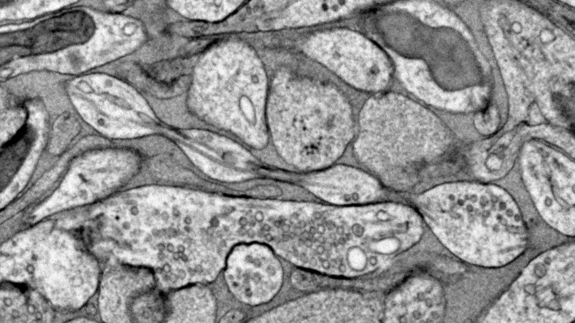
The ultrathin tendrils that zip messages around the nervous system are often drawn as smooth lines. But a new study adds some flourishes to that classical picture. Like a garland of cranberries hung on a Christmas tree, nerve fibers called axons may actually be a series of bumps, researchers suggest December 2 in Nature Neuroscience.
The claim is intriguing, but it’s too soon to redraw axons, says Pramod Pullarkat, a physicist who studies neuron structure at Raman Research Institute in Bengaluru, India. “I would not take this as an absolute fact yet,” he says. “But it is very much a possibility.”
Physicists who study fluids have known about this sort of pearls-on-a-string shape for a long time. Thin strands of fluids form beads when stretched, a phenomenon that can be spotted in viscous fluids like honey and aloe vera gel.
Beads have also been spotted on axons, which are in some ways like those liquids, with malleable internal and external parts. But the beaded structure hasn’t been systematically studied.
In the new work, researchers scrutinized axons from mouse brains. Scientists commonly use chemicals to preserve or “fix” cells so they can be studied further. But that method often changes the tissue’s shape, like desiccating a grape into a raisin, says Shigeki Watanabe, a cell biologist and neuroscientist at Johns Hopkins University School of Medicine. To see axons as they truly are, Watanabe and colleagues used a high-pressure freezing method instead. This sort of cryopreservation “is like making a frozen grape,” he says.
These frozen mouse axons were made of rotund blobs connected by thin tubes, electron microscope images revealed. The researchers did the experiments with a type of axon that isn’t wrapped in insulating material called myelin. Like a puffy coat, myelin covers some nerve fibers in the brain called white matter. Further unpublished experiments show a similar pearls-on-a-string structure on myelinated axons, Watanabe says. And his team has seen pearling in human axons.
Pullarkat points out that there are clear examples of smooth axons. “People have been looking at axons for a long time,” he says. Cells grown in dishes, for instance, usually have thick axons that look cylindrical unless they’re damaged. “But it is possible that a subset of axons have this pearl geometry even in normal conditions.”
These blobs — formally known as nanoscopic varicosities — form because of physical mechanics, Watanabe says. It takes less energy to make the blob structure than it does to make a cylinder, he says.
An axon’s shape influences how fast signals move along it, modeling experiments and experiments in mice axons suggest. And there are hints from the new study that signals moving along the axons influence the shape of the bumps, too.
But the matter isn’t settled. The freezing method could be distorting the axons somehow. “It’s possible that the technique is not there yet, to actually see things properly,” Watanabe says. “But I feel confident about what we observe.”
Watanabe and colleagues plan to study whether pearled axons are affected by sleep, a time when the mechanical environment changes in the brain. They’re also eager to study the shapes more by looking at axons inside living brains (SN: 11/9/17).
Pullarkat is happy to see the data, whether it turns out to be a general axon shape or a phenomenon that is more selective. “Sometimes it’s not easy to shake up an established fact, and people just sort of hesitate do it,” he says. In the case of the pearling axons, “there is good enough reason to think that maybe they are onto something which is real.”
So stay tuned — but don’t take a Sharpie to the axon drawing just yet.
Source link
