New Tools For Old Diagnosis
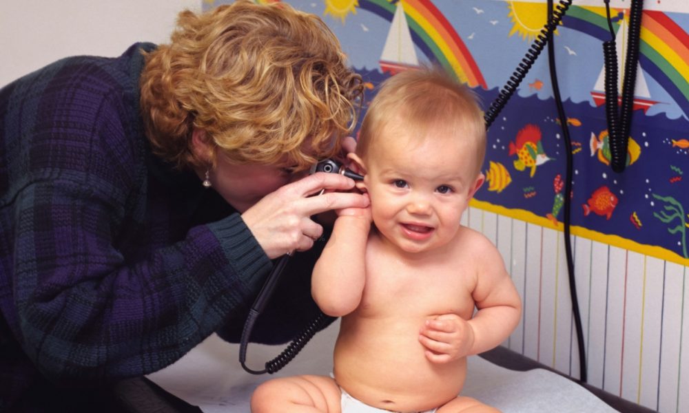
Since the dawn of medicine, many diagnostics rely on the visual inspection of a patient. This is still true for ear infections, usually requiring an experienced clinician to be properly diagnosed.
In this specific case, the experience and skill of the doctor matter a lot, as previous studies of clinicians have reported diagnostic accuracy of AOM ranging from 30% to 84%, depending on type of health care provider, level of training, and age of the children being examined.
As a result, many ear infections are misdiagnosed as otitis media with effusion, or fluid behind the ear, a condition that generally does not involve bacteria and does not benefit from antimicrobial treatment.
But a new tool has now been developed to help doctors, relying on the emergence of AI machine vision, together with the omnipresence of smartphones. And it could help reduce unnecessary use of antibiotics, which leads to the problematic emergence of antibiotic resistance.
“Underdiagnosis results in inadequate care and overdiagnosis results in unnecessary antibiotic treatment, which can compromise the effectiveness of currently available antibiotics. Our tool helps get the correct diagnosis and guide the right treatment.”
Alejandro Hoberman, M.D., Pr. of pediatrics and director of the Division of General Academic Pediatrics at Pitt’s School of Medicine.
Source: UPMC
It is also a very common problem, with 70% of children catching an ear infection in their first year of life.
AI Diagnosis
Pr Hoberman teamed up with researchers at Tandon School of Engineering in NY, Bosch Center for Artificial Intelligence in Pittsburgh, and Dcipher Analytics in Stockholm, Sweden to develop an AI tool for ear infection detection.
They published their findings in JAMA Pediatrics, under the title “Development and Validation of an Automated Classifier to Diagnose Acute Otitis Media in Children.
The resulting AI tool only requires an otoscope connected to a smartphone camera.
They created two different AI models and used a database of 1,151 videos of the tympanic membrane from 635 children. They then asked experts to manually annotate each of the videos as corresponding to an ear infection or not.
921 videos were used for training the AI, and the leftover 230 videos were used as a test to assess the AIs” accuracy.
Among the parameters measured for correct diagnostics were the shape, color, position, and translucency of the eardrum.

Source: UPMC
Superior Medical Results
Both models were highly accurate, producing a sensitivity of 9.38% and a specificity of 93.5%. This means that not only did the AIs detect the infection accurately, but they also had a very low rate of false negatives and false positives.
It is also worth noticing that this is a better accuracy than even the best results obtained by visual identification by doctors, and much higher than the results obtained in less ideal conditions (younger children, untrained doctors, etc.)
The video can be recorded and archived. This first can be used to explain the diagnosis to the patient or the patient’s parents and be archived in the patient’s file. The recorded videos can also be used to train medical students or residents and provide a valuable teaching tool for hospitals and medical practices.
It should also help family doctors to make the right diagnosis and reduce the overprescription of antibiotics.
The simplicity of the implementation of this tool, using only a doctor’s smartphone and an otoscope should also allow for a very quick deployment and ease of adoption.
AI Diagnostic Companies
For a long time, applying AI to complex environments like the human body was not working, as it struggled to deal with the “messiness” of the data provided.
New technologies like neural networks have changed this, creating “machine vision” of which some of the more well-known applications are self-driving cars.
A lot of medical diagnoses today still rely on the opinion of doctors and the expertise coming from studying manually thousands of images of scanners, RMI, and also eardrums. If AIs now can determine where cyclists are on the road, they are also getting better, if not better than humans, at detecting infections, tumors, and other medical issues.
Many large companies are incorporating AI into their imaging systems, like GE Healthcare, Siemens Healthineers, Canon Medical, and Philips. However due to the size of the corporations they are hardly pure players in AI medical diagnostics.
Other more focused startups are privately listed, like PathAI and Viz.AI for example. So we focused on publicly traded stocks instead.
1. Butterfly Network
Butterfly is both the developer of an advanced ultra-portable ultrasound diagnosis tool and an integrated software using AI helping diagnosis, called “Compass”.

Source: Butterfly Network
The company is now on its 3rd generation of the ultrasound probe, with the release in 2024 of the iQ3, with a higher data transfer rate and 2x the processing power of the version before it. Like all previous Butterfly ultrasound probes, it relies on the superior semiconductor “ultrasound-on-chip” technology instead of classical piezoelectric sensors.
iQ3 offers a superior user experience, including the possibility to visualize both 3D and multi-planes simultaneously, integrated cloud software, and quick start-up, all for a cheaper price.
The company uses AI to improve the images, generate diagnosis-relevant measurements automatically, as well as provide training/teaching practice.

Source: Butterfly Network
Butterfly is quickly expanding into new markets in Asia (Singapore, Indonesia, Philippines, etc.), and also in the veterinarian markets, for example, checking feedlot cattle health, leveraging the ultra-portability of its ultrasound tool.
2. Enlitic (ENL.AX)
Enlitic has developed an AI system to analyze radiology images and automatically generate standardized descriptions of the images. So this represents the next step in radiology images big data, following the adoption of technical standards like DICOM or HL7.
To do so, Enlitic’s technology utilizes computer vision and natural language processing to analyze DICOM images, identifying various parameters like body parts, orientation, contrast, and slice thickness for CT, MR, and X-ray images.

Source: Enlitic
This sort of standardization will be required for the progress of AI in radiology, as well as telemedicine and automation. It will provide interoperability between systems, depending on a reliable labeling system.
It should also help monetization of this data by hospitals and radiology centers, to provide anonymized and standardized data for other AI training.
